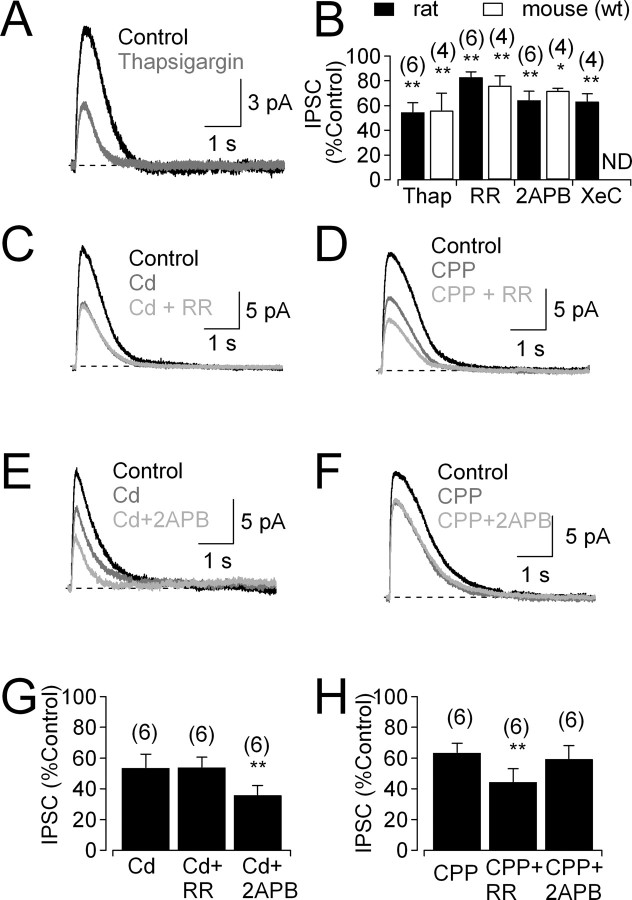
Figure 7.
IP3Rs and RyRs contribute to CICR in glycinergic amacrine cells. A, Feedback IPSC before and after bath application of thapsigargin (1 μm). B, Summarized effects (mean ± SD) of thapsigargin (1 μm), RR (40 μm), 2-APB (50 μm), and xestospongin C (XeC; 3 μm) on IPSCs. ND, Experiment not done. C, IPSCs recorded under control conditions, in the presence of Cd2+ and the additional presence of RR. D, IPSCs recorded under control conditions, in the presence of CPP and the additional presence of the RR. E, F, As in C and D, but with the IP3R antagonist 2-APB. G, H, Summarized data (mean ± SD) showing that IP3Rs and RyRs amplify specifically NMDAR-mediated and Cav channel-mediated Ca2+ signals, respectively. All experiments were performed in the presence of GABAR antagonists (SR95531 and TPMPA).

