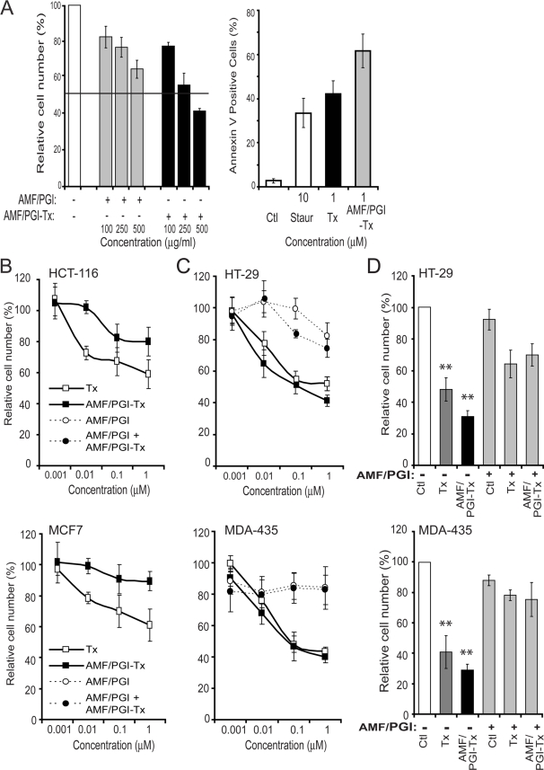
Figure 3. The growth inhibition and pro-apoptotic effects of AMF/PGI-paclitaxel.
A. The specificity of AMF/PGI-paclitaxel was determined by competitive binding assay. MDA-435 cells were incubated for 60 min at 4°C in the presence of various concentrations (100–500 µg/ml) of either free AMF/PGI or AMF/PGI-paclitaxel conjugate. Afterwards, cells were stained with anti-gp78/AMFR mAb (3F3A) and Alexa647-conjugated secondary antibody and surface expression of gp78/AMFR determined by flow cytometry (left panel). Results were normalized and expressed as means±SE (n = 4) compared to the control (untreated cells). Induction of apoptosis by 10 µM staurosporine, 1 µM paclitaxel (Tx) or 1 µM AMF/PGI-paclitaxel conjugate was determined as described in the Material and Methods by flow cytometry using the Annexin V–FITC assay (right panel). The growth inhibitory ability (B, C) and selectivity (D) of AMF/PGI-Paclitaxel conjugate were assessed on HCT116 and MCF7 cells that poorly internalize AMF/PGI (B) and HT29 and MDA-435 cells that efficiently internalize AMF/PGI (C, D). Cells were treated with increasing log concentrations of paclitaxel equivalent concentrations (0–1 µM) of free paclitaxel, AMF/PGI-paclitaxel conjugate, or controls, as indicated, for 48 hours (B, C). Alternatively, HT29 and MDA-435 cells were treated with 1 µM free paclitaxel or AMF/PGI-paclitaxel in the presence or absence of a 20× fold excess of unconjugated AMF/PGI for 48 hours (D). Cell viability was then measured using crystal violet staining. Each measurement was done in quadruplicate and the results are presented relative to untreated control cells. Results were normalized and expressed as mean±SE (n = 4) compared to the control (untreated cells), **, P≤0.001 versus control.

