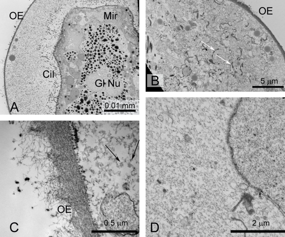
Figure 6. Miracidia of S. japonicum, fixed immediately after shell rupture for conventional processing for TEM.
A. Low magnification image showing miracidium within the expanded lacuna. The penetration glands are evident in the miracidium. B. Expanded lacuna with numerous membranes (arrows). C. Outer envelope. Note that the external fibrils appear to be unravelling. Partly degraded rosettes are shown (arrows). D. Lacuna, showing membrane body, presumably a vacuole. Cil- cilia; Gl Nu- nuclei of penetration glands; Mir- miracidium; OE- outer envelope.

