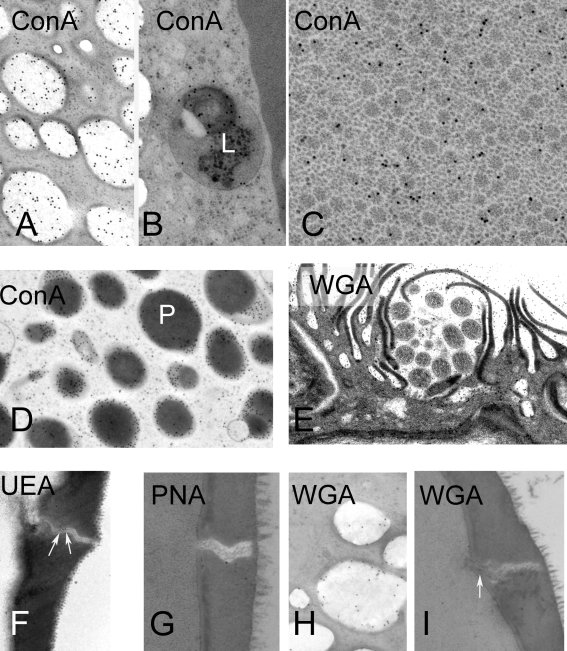
Figure 7. Lectin cytochemistry of S. japonicum eggs, shown by TEM, prepared by HPF and cryo-substitution.
The panels show representative regions with positive localization with the lectin. A–D Con A cytochemistry. A. Lipoid bodies B. Lysosome in inner envelope. C. Rosettes in vacuoles; D. Penetration gland of miracidium. E. WGA labelling in lacuna surrounding the terebratorium. F. UEA in pore of shell. G. PNA in pore of shell. H. WGA in lipoid body. I. WGA in pore.

