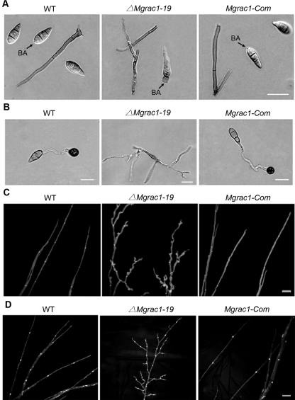
Figure 2. Abnormal conidial morphology, appressorial formation, and hyphal branching in the Mgrac1 deletion mutant.
(A) Differential interference contrast (DIC) microscopy of conidia cultured on an oatmeal agar plate at day 10 after incubation. BA = basal appendage where conidia attach to conidiophores. Bar = 20 µm. (B) Conidia incubated on the surface of artificial hydrophobic Gelbond films as described in Materials and Methods. Bar = 20 µm. (C) Branching patterns of mycelia on complete media plates at day 3 after incubation. Frequent branching occurs at the terminal mycelia of ΔMgrac1-19. Calcofluor staining of mycelia is used to show the distance of septa. Bar = 20 µm. (D) DAPI staining of mycelia to show the localization of nuclei. Bar = 20 µm.

