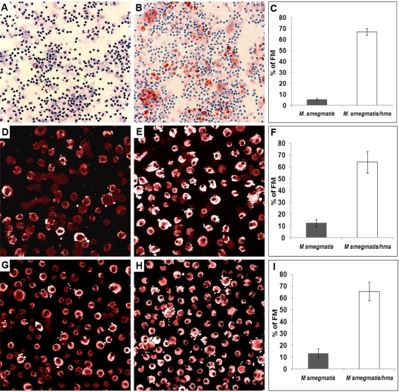
Figure 4. Role of hma-controlled mycolic acids in FM formation.
PBMCs from a healthy donor were infected with either M. smegmatis or M. smegmatis/hma for 3 days. The granuloma cells were then collected and stained with Oil red-O and May-Grünwald Giemsa (A, B). In parallel experiments, isolated macrophages were infected with either M. smegmatis (D) or M. smegmatis-hma (E), or incubated with mycolic acids extracted from both species (M. smegmatis G, M. smegmatis-hma H). The cells were then stained with Nile red. The pictures are representative of 3 independent experiments with 3 unrelated controls. Original magnification: A, B: ×100; D, E, G, H: ×400. The percentage of FMs in the different macrophage populations are indicated in panels C, F, I, respectively.

