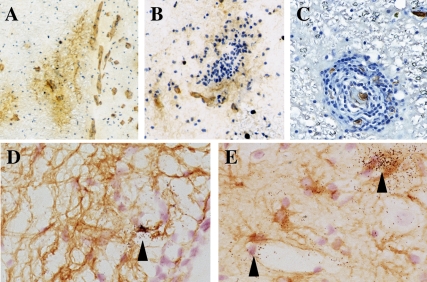
Figure 2. Detection of Glut-1 and HTLV-1 transcripts in the thoracic spinal cord from HAM/TSP patient.
(A,B,C) Glut-1 staining by iPO (DAB substrate, brown) on thoracic spinal cord sections from a HAM/TSP patient. Nuclei were counterstained with Harri's haematoxilin solution (blue). (A,B) Cryostat section; (C) paraffin section. Magnification: (A) 30×; (B,C) 75×. (D,E) Detection of HTLV-1 transcripts (tax) in cryosections of the spinal cord by in situ hybridization. Arrow heads indicate positive cells and vascular structures. Astrocytes were detected by iPO (DAB substrate, brown) against GFAP. Magnification: 220×.

