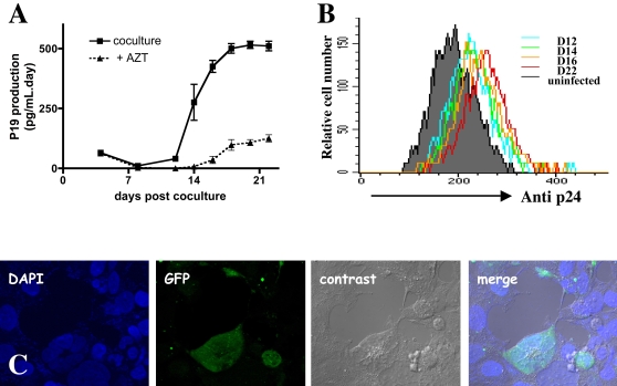
Figure 5. Productive infection of hCMEC/D3 cells by HTLV-1.
(A) Kinetics of p19 viral protein secretion in the supernatant of hCMEC/D3 and irradiated HTLV-1 MT-2 lymphocyte cocultures. Cells were cultivated or not in presence of 25 µM AZT. Results are mean and standard deviation from triplicate experiments. (B) Kinetics of detection of infected hCMEC/D3 cells by irradiated MT-2 cells. Infection was assessed by FACS analysis of p24 viral protein at days 12, 14, 16 and 22 post-coculture. (C) Production of infectious viral particles by HTLV-1-infected endothelial cells using a reporter cell-line (293T-LTR-GFP). The supernatant of hCMEC/D3 infected cells was collected and ultracentrifuged and the resuspended pellet was applied on the reporter cell-line 293T-LTR-GFP. The expression of GFP was assessed 6 days later after cell fixation.

