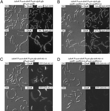
Fig. 4.
DivK controls the polar localization of ClpX and CtrA. (A and B) DIC and fluorescence images of ClpX-GFP in the presence of CpdR or CpdRD51A, and in the presence or absence of DivK. Cultures from the indicated strains were grown in M2G medium with vanillate, to induce divK expression, and aliquots were incubated in either the presence or absence of vanillate. After 8 h of DivK depletion, cells were induced with xylose (for clpX-gfp and cpdR or cpdRD51A expression) for 2 h. Proportion of cells showing at least one polar ClpX-GFP fluorescence focus (white arrows) and number of cells observed are indicated. (C and D) DIC and fluorescence images of YFP-CtrA-RD-15 in the presence of CpdR or CpdRD51A, and in the presence or absence of DivK. After 8 h of DivK depletion, cells from the indicated strains were induced with xylose (for yfp-ctrA-RD-15 and cpdR or cpdRD51A expression) for 2 h. Polar YFP-CtrA-RD-15 fluorescence foci are indicated (white arrows).

