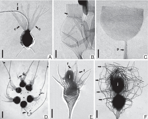
Fig. 1.
Morphological variation within choanoflagellates. Shadowcast whole mounts of cells or thecae viewed with transmission electron microscopy. (A) Monosiga ovata. (c) collar; f, flagellum. Bar = 2 μm. (B) Salpingoeca urceolata. Empty flask-shaped theca is shown. Arrow denotes inner flange that connects cell (absent) to theca. (Scale bar, 1 μm.) (C) Salpingoeca infusionum. Empty cup-shaped organic theca is shown. (p) peduncle (stalk). (Scale bar, 1 μm.) (D) Salpingoeca amphoridium. Colonial “proterospongia” stage is shown. Note six regularly placed cells held together by fine posterior threads. (Scale bar, 5 μm.) (E) Acanthoeca spectabilis. Immediately after division (nudiform) showing two cells, each with a forwardly directed flagellum (arrows in F) is shown. The juvenile (j) is above the cell remaining in the parent lorica. (Scale bar, 2 μm.) (F) Stephanoeca diplocostata. Tectiform division showing inverted juvenile cell (j) being pushed into an accumulation of costal stripsis shown. Arrows denote transverse (ring) costae. (Scale bar, 2 μm.)

