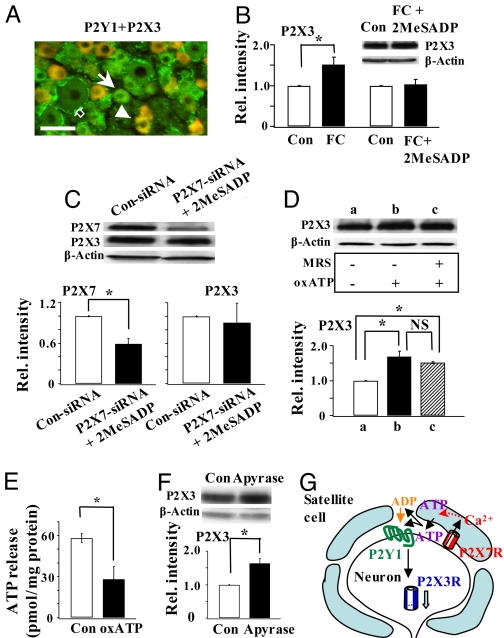
Fig. 3.
P2Y1R activation is necessary and sufficient for the regulation of P2X3R expression by P2X7Rs. (A) P2Y1Rs were co-expressed with P2X3Rs in a number of small DRG neurons (arrowhead). Some neurons expressed only P2Y1Rs (arrow). Only a few satellite cells were found to express P2Y1Rs (empty arrow). Bar = 50 μm. (B) P2X3R expression increased after FC treatment (FC/control = 1.52 ± 0.18, n = 3, *P < 0.05; data were re-plotted from Fig. 1B). In the presence of 2MeSADP, FC could no longer change P2X3R expression ([FC + 2MeSADP]/control:1.03 ± 0.12, n = 3, P > 0.05). (C) Activation of P2Y1R is sufficient for the inhibitory control of P2X3R expression. Down-regulation of P2X7Rs no longer elicited an increase in P2X3R expression when rats were treated with 2MeSADP (P2X7R: 0.59 ± 0.08, n = 3, *P < 0.05; P2X3R: 0.9 ± 0.29, n = 3, P > 0.05). (D) In the presence of oxATP, P2X3R expression was up-regulated (oxATP/control 1.68 ± 0.16, *P < 0.05, n = 2). Blocking the activity of P2Y1R by MRS could not further affect the expression of P2X3Rs ([MRS + oxATP]/control: 1.51 ± 0.03, *P < 0.05, n = 2; oxATP/[MRS + oxATP] = 1.11 ± 0.09, n = 2, P > 0.05). (E) P2X7Rs are involved in the basal ATP release. Basal ATP release was much reduced when P2X7R activity was blocked by oxATP (control: 58.03 ± 3.56 pmol/mg protein, oxATP: 28.13 ± 9.16 pmol/mg, n = 3, *P < 0.05). (F) Degrading ATP and ADP by apyrase (20 U/ml) significantly relieved the inhibitory control of P2X3R expression (apyrase/control: 1.64 ± 0.15, n = 3,*P < 0.05). (G) Schematic shows the involvement of P2Y1Rs in P2X7R–P2X3R inhibitory control.

