Figure 3.
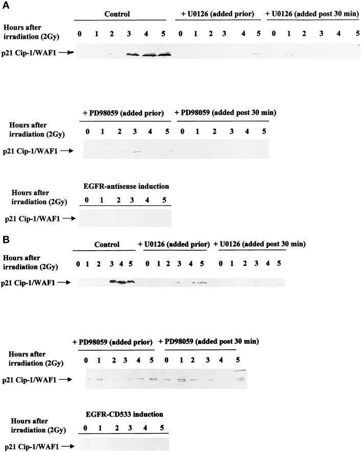
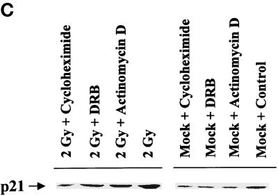
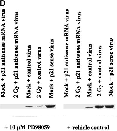
Radiation-induced p21 expression in A431-TR25-EGFR-AS cells and MDA-TR15-EGFR-CD533 cells is dependent on EGFR function and the second phase of MAPK activation. A431-TR25-EGFR-AS cells (A) and MDA-TR15-EGFR-CD533 cells (B) were either treated with doxycycline 24 h before irradiation or pretreated for 30 min before irradiation with the MEK1/2 inhibitor PD98059 (10 μM) or U0126 (2 μM). In parallel, other plates of cells were also treated 30 min after irradiation with the MEK1/2 inhibitor PD98059 or U0126. Cells were irradiated (2 Gy), and the protein expression of p21 was determined from 0 to 300 min. Cells were lysed, and equal portions (∼100 μg) from each plate were subjected to SDS-PAGE, followed by immunoblotting to determine p21 protein levels. Ponceau S staining revealed equal protein loading on the nitrocellulose (our unpublished results). Exposure was for 1 min. A representative experiment is shown (n = 3). (C) A431-TR25-EGFR-AS cells were infected with a control adenovirus to express β-galactosidase, a virus to express antisense p21 mRNA, or a virus to express p21 protein. Twenty-four hours after infection, cells were pretreated for 30 min before irradiation with PD98059 (10 μM) orvehicle control (DMSO). Cells were irradiated (2 Gy) or mock exposed, and the protein expression of p21 was determined after 4 h. Cells were lysed, and portions (∼400 μg) from each plate were subjected to SDS-PAGE, followed by immunoblotting to determine p21 protein levels. Ponceau S staining revealed equal protein loading on the nitrocellulose (our unpublished results). Exposure was for 3 min. A representative experiment for each condition is shown (n = 5). (D) A431-TR25-EGFR-AS cells were pretreated for 5 min before irradiation with DRB (30 μM), actinomycin D (5 μM), or cycloheximide (20 μg/ml) for 4 h. Cells were irradiated (2 Gy) or mock exposed, and the protein expression of p21 was determined after 4 h. Cells were lysed, and portions (∼400 μg) from each plate were subjected to SDS-PAGE, followed by immunoblotting to determine p21 protein levels. Exposure was for 3 min. A representative experiment for each condition is shown (n = 5).


