
Figure 2.
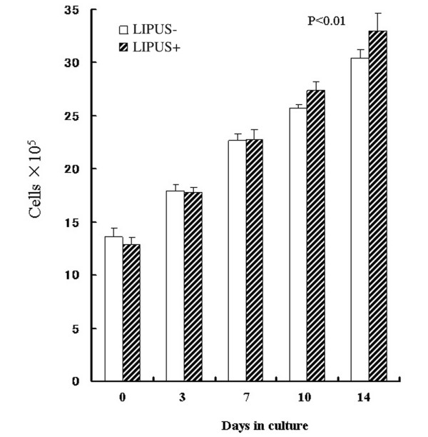
Growth curves of the cells in the chondrocyte–collagen sponges (n = 7). A time-dependent increase in the number of chondrocytes can be seen in both the low-intensity pulsed ultrasound (LIPUS) group (US+) and in the control group (US-). The rate of increase in the chondrocytes number was significantly greater, however, in the LIPUS group in comparison with the control group (P < 0.01). The change in the number of chondrocytes was assessed using repeated-measures analysis of variance.