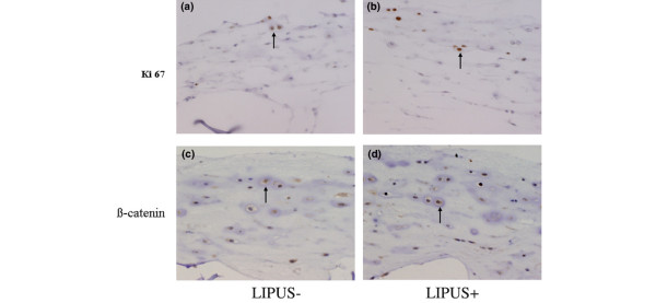
Figure 4.
Ki67 and β-catenin antibody staining. (a), (b) Anti-Ki67 antibody staining of week 2 cultures (200× magnification). The nuclei are positively stained with an anti-Ki67 antibody in both the control group (US-) and the low-intensity pulsed ultrasound (LIPUS) group (US+) (black arrows). (c), (d) Anti-β-catenin antibody staining of week 2 cultures (200× magnification). The nuclei are positively stained with an anti-β-catenin antibody in both the control group (US-) and the LIPUS group (US+) (black arrows).

