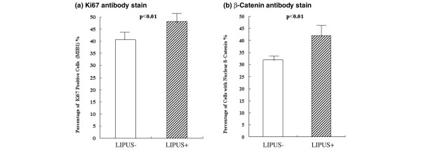
Figure 5.
Quantitative evaluation of Ki67-positive cells and β-catenin-positive cells. After counting 100 cells in each specimen in the low-intensity pulsed ultrasound (LIPUS) group (US+) and the control group (US-), the numbers of cells with positively stained nuclei were compared for both (a) Ki67 and (b) β-catenin. There were significantly more brown stained cells in the LIPUS group in both cases (P < 0.01).

