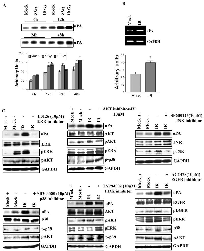
Figure 1. Radiation increases uPA and other signaling pathway molecules expression in IOMM-Lee cells.
(A) Fibrinogen zymography using the conditioned medium from IOMM-Lee cell cultures radiated with 5 or 10Gy and controls for the indicated time points. Equal amounts of total protein (0.5μg) were resolved for each sample. Mean densitometric values ± SEM were calculated and plotted as a histogram. *Statistically different compared to respective control and radiated groups (p<0.05). (B) RT-PCR analysis for uPA expression. Total RNA was extracted from IOMM-Lee cells incubated in serum-free medium for 6h after cells were radiated (with 10Gy) or not irradiated (mock). GAPDH was used as a loading control. Mean densitometric values ± SEM were calculated and plotted as a histogram. *Statistically different compared to control and radiated groups (p<0.05). (C) EGFR, through ERK1/2 and p38 and to a lesser extent, pI3K/AKT, mediates radiation-induced uPA in IOMM-Lee cells. Fibrinogen zymography of conditioned medium, and western blot analysis of cell lysates from IOMM-Lee cells. Cells were treated with specific inhibitors of JNK (SP600125) (15μM), ERK1/2 (U0126) (10μM), p38 (SB203580) (10μM), AKT (AKT inhibitor IV) (10μM), pI3K (LY294002) (20μM) and EGFR (AG1478) (10μM) for 30 min before they were radiated with 10Gy and subsequently incubated for 6h in serum-free medium. Conditioned medium was collected and 0.5μg of total protein was used for fibrinogen zymography to detect uPA activity. Equal amounts of total proteins from cell extracts were resolved by SDS-PAGE and probed with antibodies against phosphorylated JNK, ERK1/2, p38, AKT, EGFR and total JNK1/2, ERK1/2, p38, AKT and EGFR. GAPDH was used as a loading control. Experiments were repeated at least three times.

