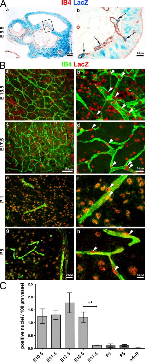
Figure 1.
Canonical Wnt signaling is active in ECs during brain angiogenesis and becomes progressively down-regulated during vessel maturation. (A) LacZ whole-mount stained (blue) E9.5 BAT-Gal embryos sectioned and counterstained for isolectin B4 (IB4; red). Panel b is a higher magnification of the boxed area in panel a. Arrows point to nuclear LacZ reporter gene staining. (B, a–d) Whole-mount hindbrain staining for LacZ (reflection; red) and IB4 (green) of BAT-Gal embryos (E13.5 and 17.5) analyzed by confocal microscopy. Arrowheads indicate LacZ-positive nuclei. (B, e–h) Staining of brain cryosections from postnatal BAT-Gal pups (P1 and P5) for LacZ (immunofluorescence [IF], red) and IB4 (green). Arrowheads indicate LacZ-positive nuclei. Positive nuclei outside the vascular system indicate active Wnt signaling in the brain parenchyma. (C) Quantification of LacZ-positive nuclei per 100-μm vessel length shows a significant decrease from E15.5 to 17.5 (five fields per hindbrain; three brains; **, P = 0.0003). Error bars represent SEM.

