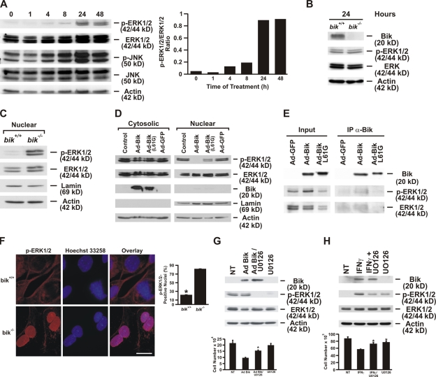
Figure 4.
Bik binds to activated ERK1/2 and inhibits its nuclear translocation. (A) HBEC-2 cells were treated with 50 ng/ml human recombinant IFNγ, and ERK1/2 activation was assessed at 1, 4, 8, 24, and 48 h of IFNγ treatment. The ratio of phospho-ERK1/2 to total ERK1/2 at different time points after IFNγ treatment was quantified. (B) ERK1/2 activation in MAECs from bik+/+ mice compared with those from bik−/− mice after IFNγ treatment. MAECs were treated with 50 ng/ml murine recombinant IFNγ for 24 h, and protein extracts were analyzed for Bik expression and phospho-ERK1/2. (C) Nuclear extract from IFNγ-treated bik+/+ and bik−/− MAECs was analyzed for activated ERK1/2. (D) Translocation of phospho-ERK1/2 is inhibited by Bik expression in primary HAECs. HAECs were either left untreated or were infected with Ad-Bik, Ad-BikL61G, or Ad-GFP at an MOI of 100, and ERK1/2 was activated by maintaining cells in unchanged media for 48 h. Nuclear and cytosolic extracts were analyzed for phospho-ERK1/2, total ERK1/2, Bik, lamin, and actin. The figure is representative of three independent experiments. (E) Bik interacts with phospho-ERK1/2. Cell lysates prepared from HBEC-2 cells infected with either Ad-GFP, Ad-Bik, or Ad-BikL61G were immunoprecipitated with anti-Bik antibody. The cell lysates (input) and immunoprecipitates were resolved by SDS-PAGE and analyzed by Western blotting using antibodies to Bik, phospho-ERK1/2, and total ERK1/2. (F) Representative photomicrographs and quantification showing that a higher percentage of MAECs from bik−/− compared with bik+/+ mice displays nuclear localization of activated ERK1/2. MAECs were treated with 50 ng/ml IFNγ for 24 h, fixed in paraformaldehyde, and immunostained for phospho-ERK1/2. The percentage of nuclei with phospho-ERK was quantified from three independent experiments. Bar, 10 μm. (G) HBEC-2 cells were infected with 100 MOI Ad-Bik and were cotreated with 1 μM U0126, the ERK-specific inhibitor. Cells infected with Ad-Bik showed a 60% decline in total cell number, and this decline was diminished when the HBEC-2 cells were cotreated with 0.1 or 1 μM U0126. The corresponding Western blot of proteins extracted from cells infected with Ad-Bik and treated with U0126 to suppress ERK1/2 activation. (H) Cell counts and Western blot analysis of protein extracted from HBEC-2 cells that were left untreated or treated with IFNγ either alone or with U0126. Treatment with 1 μM U0126 did not affect cell growth. NT, not treated. Error bars indicate group means ± SEM (n = 4 different treatments per group). *, P < 0.05; significantly different from Ad-Bik–infected cells.

