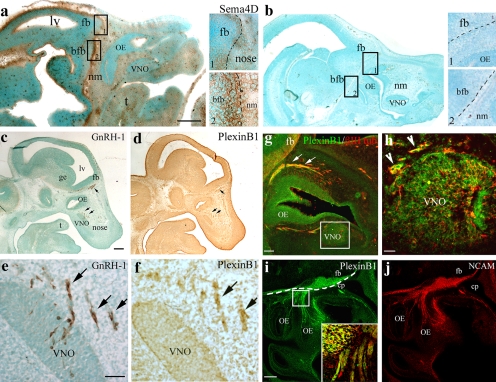
Figure 1.
Sema4D and PlexinB1 are expressed along the GnRH-1 migratory route. (a) Sema4D immunoreactivity is detected in the nasal mesenchyme (nm), surrounding the OE and VNO epithelia. A robust staining is evident at the nasal forebrain junction and rostral forebrain (see higher magnification view of box 1) as well as in the basal forebrain (bfb; see higher magnification view of box 2). (b) The specificity of the staining was confirmed by omission of the primary antibody incubation (see high-power view images in boxes 1 and 2). Sections in panels a and b were counterstained with the nuclear dye methyl green to allow visualization of the background tissue. (c–f) Consecutive sagittal sections of an E12.5 mouse immunostained, respectively, for GnRH-1 and PlexinB1. (c and e) GnRH-1–immunoreactive cells emerged from the developing VNO and migrate through the olfactory mesenchyme (arrows) toward the forebrain (fb). GnRH-1–labeled sections were counterstained (methyl green) to allow the identification of the different anatomical structures. (d and f) PlexinB1 immunoreactivity is distributed in the developing OE, in the VNO, and in a group of cells migrating out of the presumptive VNO (see arrows in f). These cells are distributed in a spatial and temporal pattern that parallels GnRH-1 immunostaining (arrows in e and f). (g and h) A sagittal section of an E12.5 mouse nose double-stained for PlexinB1 (green) and βIII-tubulin (red) indicates coexpression of these antigens in the OE, VNO, and in cells (h, arrowheads) and fibers (g, arrows) emerging from these structures. (i and j) A sagittal section of an E12.5 mouse nose double-stained for PlexinB1 (i) and NCAM (j) indicates coexpression of these antigens along the olfactory/vomeronasal axons, as shown by high-power confocal analysis (i, inset, which is an enlarged view of the boxed region). Broken lines indicate the boundary between the nose and the forebrain. cp, cribriform plate; fb, forebrain; lv, lateral ventricle; ge, ganglionic eminence; t, tongue. Bars: (a and b) 500 μm; (a and b, insets) 125 μm; (c and d) 250 μm; (e and f) 50 μm; (g) 50 μm; (h) 30 μm; (i and j) 100 μm; (i, inset) 30 μm.

