Figure 3.
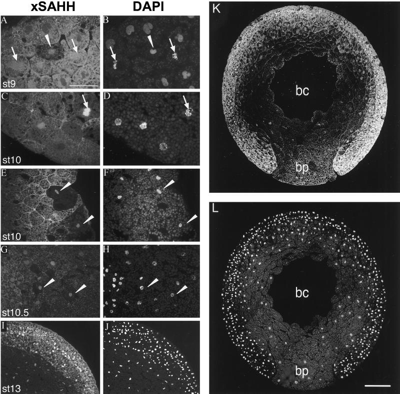
xSAHH translocates into interphase nuclei during gastrulation. Intracellular localization of endogenous xSAHH was analyzed on Technovit sections after whole-mount immunofluoresescent staining using mAb 32-5B6 (A, C, E, G, I, and K), and counterstaining of the same sections for DNA with DAPI is shown in B, D, F, H, J, and L. Details of sections cut in the animal to vegetal direction were selected that show the marginal zone of a late blastula at stage 9 (A and B), the vegetal area (C and D), and the dorsal lip of an early gastrula at stage 10 (E and F), a marginal area of a gastrula at stage 10.5, with mesoderm to the left and endoderm to the right (G and H), and all three germ layers anteriorly of a very early neurula at stage 13 (I and J). Single interphase nuclei showing nuclear xSAHH are marked with arrowheads in A–H. Mitotic cells are marked with arrows, and arrows point at prometaphase in C and D. On a complete transverse section of a gastrula at stage 11.5 (K) the endoderm of the blastoporus (bp) is flanked by the two blastoporal lips, containing mesoderm and ectoderm. bc, remnant of the blastocoel. Cytoplasmic xSAHH is excluded from yolk platelets and is concentrated at the cell boundaries. Bars: A–J, 50 μm; L, 200 μm. Sections were 2 μm thick in G and H and 5 μm thick in all other panels.

