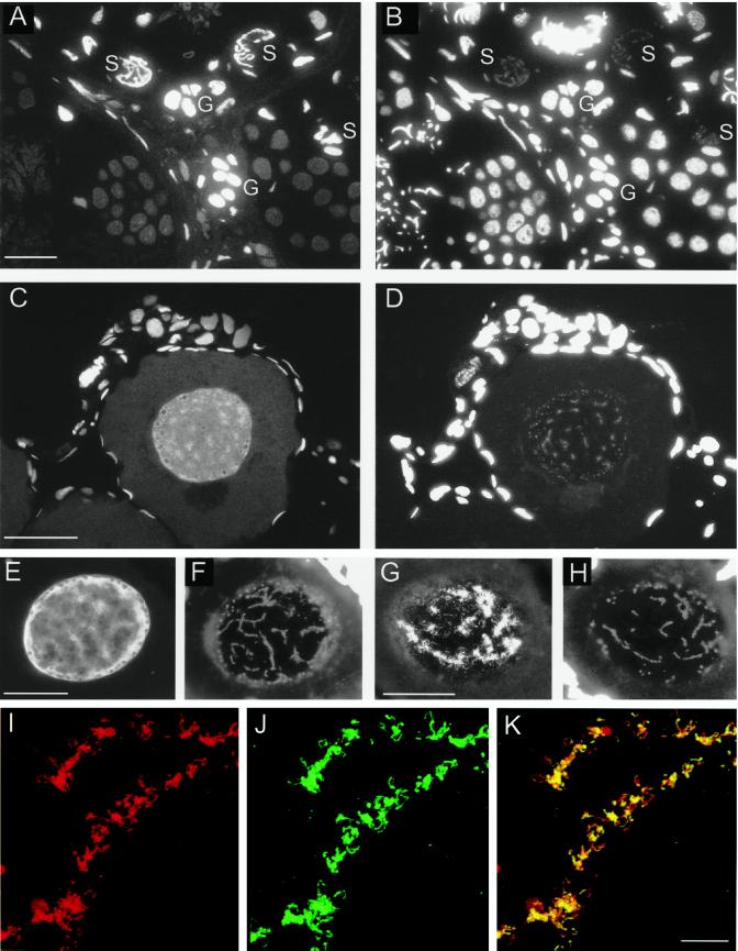
Figure 5.
Immunohistological localization of xSAHH in the nuclei of germ cells. xSAHH was detected on 1.5-μm thin sections after whole-mount immunofluorescent staining of adult testis (A) and ovary from a 4-cm-long young female (C) with mAb 32-5B6. (B and D) Counterstaining of the same sections with DAPI. In the testis (A and B), nuclear staining with 32-5B6 predominates in transcriptionally active Sertoli cells (S) and spermatogonia (G). In the ovary (C and D), xSAHH is nuclear in all cells. In the single diplotene (stage I) oocyte nucleus shown, the xSAHH antigen appears most highly concentrated around the lampbrush chromosomes, which are weakly stained with DAPI in D. (E–H) Frozen sections of an ovary were stained with mAb 32-5B6 (E) and with anti-RNA Polymerase II mAb H14 (G). (F and H) DAPI counterstain of the sections shown in E and G. Follicle cell nuclei stained with DAPI are overexposed in D, F, and H to visualize lampbrush chromosomes. Bar, 50 μm. (I–K) Lampbrush chromosome spread from a stage V oocyte double stained for xSAHH (I) and for RNA polymerase II (J). The confocal images shown in I and J are superimposed in K. Bar in K, 10 μm.

