
Figure 4.
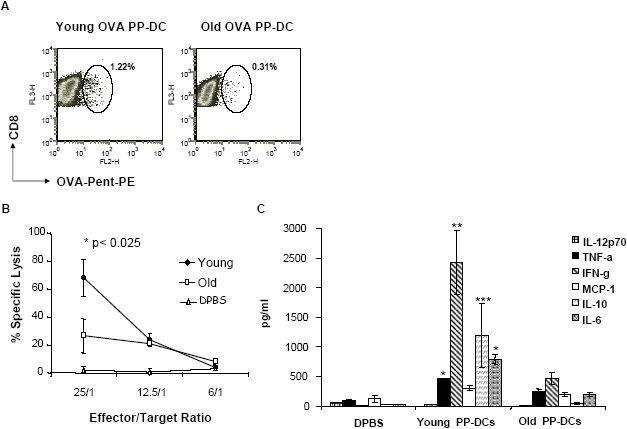
The effect of young and old DCs on T cell functions. (A) In vivo detection of OVA-specific CD8+ T cells by MHC pentamer staining. Day 7 tumor-bearing B6 mice were vaccinated with young or old OVA PP-DCs. 7 days later, total splenocytes were harvested, depleted of CD19+ cells and stained for CD8-RPE Alexa Fluor 647 and OVA-Pent-PE. A representative figure of FACScan plots depicting the percentage of OVA-specific CD8+ T cells is shown. (B) CTL activity of splenocytes from mice receiving young and old DC vaccines. Spleens isolated from DC vaccinated or control mice (DPBS-treated tumor bearing mice) were stimulated for 6 days in vitro with mitomycin-C treated B16-OVA tumor cells and cytotoxic activities against B16-OVA target cells were measured. Data are expressed as mean specific lysis of quadruplicate values (%) ± standard error of the mean of 3 independent experiments. (C) Cytokine release of activated CTL cells in response to tumor stimulation. Splenocytes from vaccinated mice were co-cultured with irradiated B16-OVA cells. After 48 hrs, supernatants were collected for cytokine analysis by Cytometric Bead Arrays. Data are reported as the average amount of cytokine secreted ± SEM of 3 experiments. *p < 0.001, **p < 0.005, ***p < 0.05: young PP-DCs vs old PP-DCs.