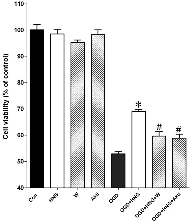
Figure 1.
The effects of HNG and the PI3K/Akt inhibitors wortmannin and Akti-1/2 on OGD-induced cell death in primary cortical neurons. OGD experiments were conducted in cultured mouse cortical neurons at DIV 10. Primary cortical neurons were incubated with 0.2 μM HNG, 0.1 μM wortmannin (W), or 1 μM Akti-1/2 (Akti) in glucose-free HBSS in a hypoxia chamber for 60 min. The plate was then restored to normoxic conditions. Cell viability was assessed by the MTS assay at 24 h of reperfusion. Control culture plates in the presence of HNG, wortmannin, or Akti-1/2 were exposed to oxygenated HBSS containing 5.5 mM glucose in normoxic condition. Bars represent mean ± SEM of 8 samples. * P<0.01 versus the OGD group; #, versus the OGD+HNG group.

