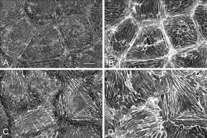
Fig. 5.
Effect of jasplakinolide treatment on blebbistatin-induced cytoskeletal alterations. No significant alterations in myosin II (A) and F-actin (B) distribution were detected after 90-min incubation with 100 nM jasplakinolide. Monolayers pretreated with jasplakinolide for 60 min and then incubated with 50 μM blebbistatin for 30 min in the presence of inhibitor show loss of organized periodic myosin II staining (C) while the majority of actin filaments remain intact (D). Bar, 10 μm.

