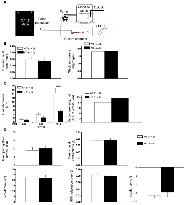
Figure 4. Muscle mechanics within isolated adult WT and Fhl1-deficient cardiac muscles following stretch.
(A) Schematic representation of isolated adult RV papillary mouse muscle from 8- to 12-week-old mice in a tissue chamber. Specimens were stretched for 5 hours to a maximum extension of 15%–20% (90%–95% Lmax). Control muscles were left in the system for the same period of time at slack length. The system was used to analyze load-inducible hypertrophic markers as well as passive and active stresses as a function of passive stretch. (B) Quantitative assessment of cross-sectional area and slack sarcomere lengths in WT and Fhl1-deficient muscles. (C) Passive tensile stress (diastolic stress) of RV papillary muscles in WT and KO muscles before and after stretch. Sarcomere lengths in WT and KO muscles at 20 kPa stress. *P < 0.05. (D) Active mechanical properties were assessed in isolated WT and KO muscles as a function of stretch.

