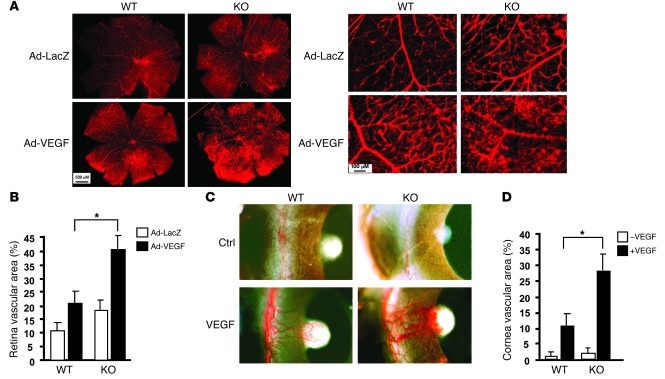
Figure 4. VEGF-induced retina and cornea neovascularization were greatly augmented in KO mice.
(A and B) VEGF-induced retina angiogenesis. Ad-VEGF or Ad-LacZ was injected intravenously into WT and KO mice. Retina vasculature was visualized by isolectin staining shown in A, with quantification of vessel density in B. (C and D) VEGF-induced cornea angiogenesis assay. A Hydron pellet containing VEGF was implanted into the cornea of WT and KO mice. Angiogenesis was assessed by stereo microscopy on day 5 following implantation (C) and vascular density was quantified in D. Data are mean ± SEM from 10 fields per tissue (ear, retina, or cornea) (n = 5 for each group). *P < 0.05. Ad-LacZ, adenoviral vector encoding the LacZ gene for expression of β-gal; Ad-VEGF, adenoviral vector encoding the VEGF gene for expression of VEGF.

