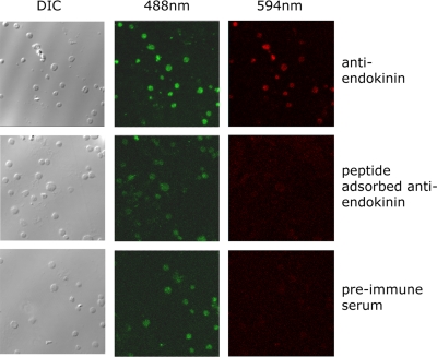
Figure 5.
EKA/B immunoreactivity is present in human platelets. Human platelets were labeled with the lipophilic fluorescent dye DIOC6, applied to glass coverslips, and fixed. Platelets were visualized by DIC microscopy and confocal microscopy at 488 nm. Endokinin expression in platelets was detected at 594 nm using an antiendokinin antibody and Texas red–conjugated secondary antibody. Parallel control experiments were performed using preimmune antiserum, and antiendokinin antiserum to which immunogen antibody had been previously added (peptide-adsorbed antiendokinin). Confocal fluorescence microscopic data represent composite images. Data are representative of 4 separate experiments.

