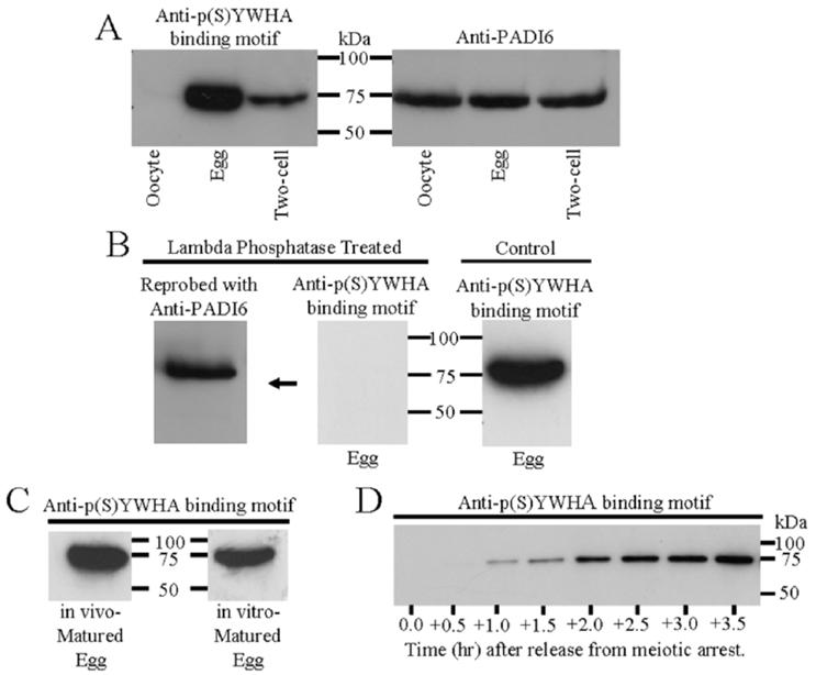
FIG. 1.

Western blots of proteins from zona-free immature oocytes, mature eggs, and two-cell embryos. Proteins were extracted, resolved by SDS-PAGE, and immunoblotted with the p(S)YWHA binding motif antibody or the PADI6 antibody following procedures outlined in Materials and Methods. All lanes were loaded with total protein extract from an equal number of cells or embryos (n 100 in A—C and 50 in D). These figures are representative of two or three repeated experiments. A) Comparative Western blots of oocytes, eggs, and two-cell embryos with the anti-p(S)YWHA binding motif or anti-PADI6 antibodies. The p(S)YWHA binding motif antibody labels a single band in eggs and two-cell embryos (but not in oocytes) that has the same molecular weight as the band labeled by the PADI6 antibody in oocytes, eggs, and embryos. No other bands were labeled by either of these antibodies. B) The p(S)YWHA binding motif antibody labels only phosphorylated proteins. No proteins are detected when a blot containing egg proteins was probed with the anti-p(S)YWHA binding motif antibody after lambda protein phosphatase treatment (center). The center blot shown was reprobed with anti-PADI6 (left) showing the presence of immunoreactive PADI6 protein at approximately 75 kDa. The control blot (right) was not treated with lambda phosphatase and was probed with the p(S)YWHA binding motif antibody showing an immunoreactive band at approximately 75 kDa. C) The p(S)YWHA binding motif antibody labels the same 75-kDa band in both in vivo- and in vitro-matured eggs. D) Phosphorylation of the 75-kDa protein begins within 1 h of release from meiotic arrest. Each lane of the Western blot was loaded with lysate from 50 prophase I oocytes (time 0) or lysate from 50 cells collected at the times indicated after the cells were washed free of dbcAMP. In this batch of cells, germinal vesicle breakdown was completed in most cells by two hours.
