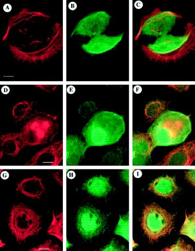
Figure 1.
ODC and cytoskeletal organization in normal human epidermal keratinocytes. To assess potential colocalization of ODC with a cytoskeletal component, NHEK were stained for ODC (B, E, or H) and actin (A), tubulin (D), or keratin (G) and optically sectioned using confocal laser-scanning microscopy, and corresponding images were superimposed to determine the degrees of overlap (C, F, or I; orange-yellow→yellow). Superposition of actin (A) with ODC (B) showed little overlap (C) of the two staining patterns. Tubulin (D) and ODC (E) staining showed little overlap (F) except in a cell undergoing mitosis in which staining was apparent throughout. Keratin (G) and ODC (H) exhibited overlap (I) in the perinuclear region of the cell with little overlap outside of that region. Digital micrographs were collected using the Noran Intervision software (Noran Instruments). Bars: A–C, D–F, and G–I, 10 μm.

