Figure 2.
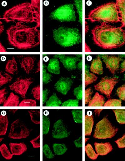
ODC and cytoskeletal organization in cytochalasin D–treated NHEK. To determine the effects of remodeling the cytoskeleton on ODC organization, NHEK were treated with 1 μg/ml cytochalasin D for 6 h, stained for ODC (B, E, or H) and actin (A), tubulin (D), or keratin (G), and optically sectioned by confocal laser-scanning microscopy, and corresponding images were superimposed to determine the degrees of overlap (C, F, or I; orange-yellow). Superposition of actin (A) with ODC (B) showed only minimal areas of overlap (C). Tubulin (D) and ODC (E) staining showed nonspecific areas of overlap (F). Keratin (G) and ODC (H) exhibited extensive overlap throughout the cells (I). Bars: A–C, D–F, and G–I, 10 μm.

