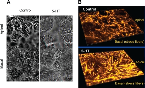
Fig. 3.
Morphological assessment of F-actin by confocal microscopy. Control Caco-2 monolayers or treated with 5-HT were stained with phalloidin. In 5-HT-treated monolayers, a dramatic reorganization of F-actin was observed such that prominent stress fibers were induced at the basal surface (A). Images were converted on 3D projections generated using Autovisualize (B). Representative results of 5–6 different experiments performed on different occasions are shown.

