Fig. 5.
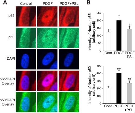
Immunofluorescent imaging shows a prednisolone-mediated suppression of PDGF-mediated nuclear translocation of p65 and p50. A: representative immunofluorescence staining of p65 and p50 in PASMC. DAPI staining was performed to indicate cell nuclei. Overlay of the images (p65 or p50 with DAPI) identifies nuclear translocation of p65 and p50 in PASMC treated with (PDGF), without (Control) PDGF (10 ng/ml), and with both PDGF and prednisolone (PDGF+PSL). B: summarized immunofluorescent staining of p65 and p50. The fluorescence intensity of p65 and p50 in the nuclear area was significantly increased in cells treated with PDGF, and concurrent treatment of the cells with PDGF and prednisolone significantly reduced the p65 and p50 in the nuclear area. All fluorescent images were taken at ×100 magnification. *P < 0.05, **P < 0.01 vs. control; #P < 0.05, ##P < 0.01 vs. PDGF bars.

