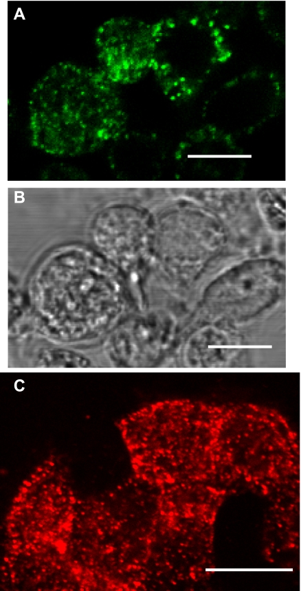
Fig. 3.
Immunolocalization of P63 in isolated rat type II cells. Type II cells were either left unpermeabilized (A and B) or were permeabilized (C) and incubated with P63 Ab. A: immunofluorescence of Alexa 488 (green)-conjugated secondary Ab. B: phase contrast microscopy of cells in A. C: immunofluorescence of Alexa 594 (red)-conjugated secondary Ab. Scale bar = 10 μm.

