Fig. 5.
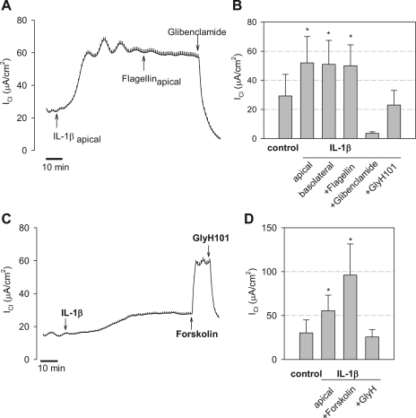
Comparisons of IL-1β and forskolin stimulation of ICl by Calu-3 cell monolayers. A: typical experiment showing effects of IL-1β (50 ng/ml, apical), P. aeruginosa flagellin (10−7 g/ml, apical), and glibenclamide (1 mM, apical+basolateral) (shown by arrows) on ICl across Calu-3 cell monolayers ([Cl−] gradient conditions). This experiment is typical of 5 others. B: averages ±SD for experiments in which IL-1β was added to the apical or basolateral surface followed by treatment with apical flagellin and either glibenclamide or GlyH101. *P < 0.05 (n = 5) for comparison to control, untreated. C: IL-1β (50 ng/ml, apical) caused characteristic, slow increases in ICl, forskolin (10 μM, apical) caused rapid increases in ICl, and GlyH101 (20 μM, apical) inhibited ICl to the control levels. D: averages ±SD for 5 experiments each identical to those shown in C. *P < 0.05 (n = 5) for comparison to control, untreated. #P < 0.05 (n = 5) for comparison of IL-1β vs. IL-1β+forskolin.

