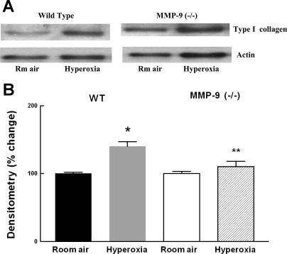
Fig. 9.
A: representative Western blot for type I collagen in 13-day-old WT and MMP-9 (−/−) mouse lungs following exposure to room air or hyperoxia for 8 days. B: densitometry of Western blots. Bars are means ± SE; n = 4. Type I collagen signal was normalized to actin. *P < 0.05 compared with room air. **P < 0.05 compared with hyperoxia-exposed WT.

