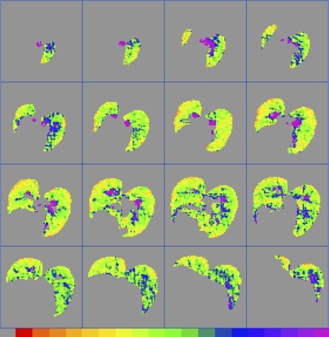
Fig. 5.
An example of a full 3D data set, taken from rat E4, showing Dave calculated on a pixel-by-pixel basis from 3He MRI data and corrected to EEP = 0 cmH2O. The left lung is on the observer's left. Planar resolution is 0.5 × 0.5 mm, and the slice thickness is 2 mm. The bottom color scale ranges from 0.0 (gray, left) to ≥2.5 cm2/s (purple, right).

