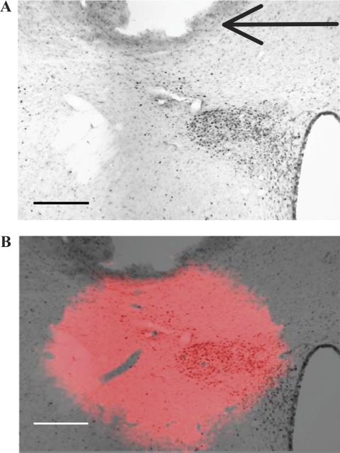
Fig. 1.
Paraventricular nucleus (PVN) cannula placement and injection spread. A: photomicrograph from a rat treated with an intra-PVN injection of 0.15 M NaCl and intravenous injection of hydralazine (HDZ), in which the brain section is immunohistochemically labeled for c-Fos protein. Following HDZ, c-Fos is expressed in the PVN. The black arrow indicates the tip of the cannula. Injection site was dorsal and lateral to the magnocellular neurons of the PVN and induced little c-Fos expression around the cannula tract. B: composite image of the overlay of the fluorescent image of 1,1′-dioctadecyl-3,3,3′,3′-tetramethylindocarbocyanine perchlorate (DiI) injection spread at the same coronal plane, Bregma −1.8 mm, as A. The spread of the intra-PVN injection is observed after aligning the cannula placement in the DiI image and c-Fos image. Scale bars are 175 μm.

