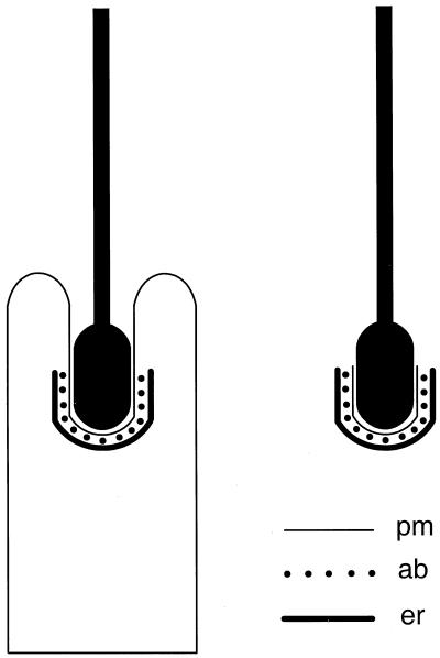
Figure 1.
Simplified schematic diagram of an ES formed between a Sertoli cell and a late spermatid in section. pm, Sertoli cell plasma membrane; er, flattened cistern of endoplasmic reticulum; ab, actin bundles. Each dot represents a parallel actin bundle cut in cross section. (Left) In the seminiferous epithelium, the head of the spermatid is held within an invagination of the Sertoli cell, and the ES is found where the Sertoli cell plasma membrane makes close contact with acrosomal region of the spermatid head. (Right) After isolation by mechanical dissociation, the majority of late spermatids retain an ES, including junctional plaque, attached to their head.

