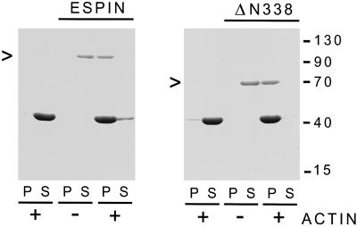
Figure 3.
Actin binding and bundling by recombinant espin and ΔN338-espin as revealed by the low-speed centrifugation assay. Shown are Coomassie blue-stained gels of the pellet (P) and supernatant (S) that result from low-speed centrifugation when rabbit skeletal muscle F-actin was incubated alone or in the presence of either recombinant full-length espin for 1 h at 4°C in 0.1 M KCl, 0.1 M imidazole-HCl, 5 mM 2-mercaptoethanol, 1 mM MgCl2, 0.5 mM ATP, 1 mM NaN3, pH 8.5 (left panel), or recombinant ΔN338-espin for 1 h at 37°C in 0.1 M KCl, 10 mM imidazole-HCl, 0.5 mM dithiothreitol, 1 mM MgCl2, 0.5 mM ATP, 1 mM NaN3, pH 7.4 (right panel). The arrowheads at the left denote the position of the recombinant espin construct. The actin is the major band migrating slightly above the 40-kDa marker.

