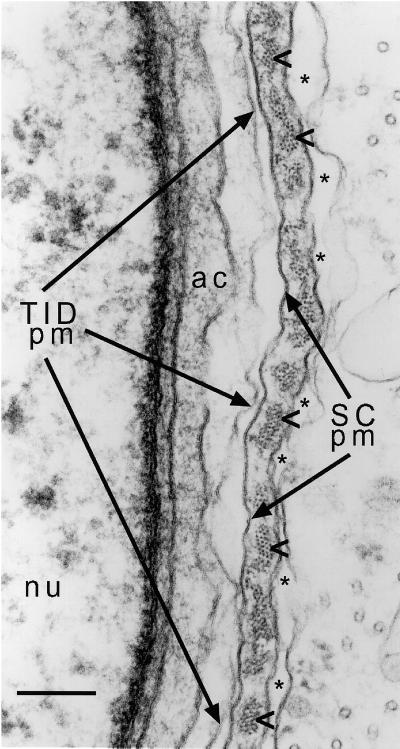
Figure 9.
Electron micrograph highlighting the various layers of the ES and neighboring structures present at the site of contact between a Sertoli cell and an early step 8 spermatid in a section of rat testis. The right portion includes the Sertoli cell and highlights its plasma membrane (SC pm) and the parallel actin bundles (arrowheads) and cistern of endoplasmic reticulum (asterisks in lumen) that comprise the ES junctional plaque. Note that the parallel actin bundles are cut in cross section or near cross section, so that each actin filament appears as a small dot. The left portion includes the spermatid and highlights its plasma membrane (TID pm), nucleus (nu), and acrosome (ac). Bar, 0.18 μm.

