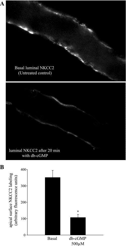
Fig. 4.
db-cGMP decreases apical surface NKCC2 staining in perfused medullary THALs. A: representative confocal micrographs showing apical surface NKCC2 staining in isolated, perfused medullary THALs under basal (unstimulated) conditions (top) and in a THAL treated with db-cGMP (500 μM) for 20 min (bottom). B: cumulative data for apical NKCC2 immunofluorescence staining under basal conditions (n = 5) and in THALs treated with db-cGMP (500 μM) (n = 6). The mean fluorescent intensity in the apical membrane was measured in 15–20 THAL cells in each tubule and then averaged.

