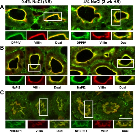
Fig. 5.
Indirect immunofluorescence microscopy of DPPIV (A), NaPi-2 (B), and NHERF-1 (C) distribution in the proximal tubules of rats on 3-wk 0.4% NaCl (NS) or 4% NaCl (HS). The kidneys were fixed as described in methods. The antibodies against DPPIV, NaPi-2, and NHERF-1 were polyclonal and detected with FITC-conjugated goat anti-rabbit secondary antibody, whereas the antibody against villin was monoclonal and detected with Alexa 568-conjugated goat anti-mouse secondary antibody. A: surface sections were double-labeled against DPPIV (green) and villin (red). B: sections were double-labeled against NaPi-2 (green) and villin (red). C: sections were double-labeled against NHERF-1 (green) and villin (red). Bar, 10 μm.

