Fig. 8.
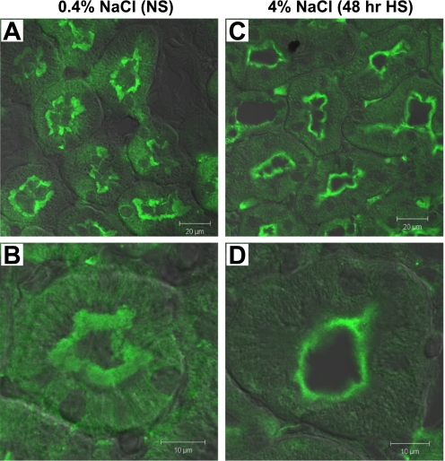
Indirect immunofluorescence microscopy and DIC overlay of AT2R in the proximal tubules of rats on 0.4% NaCl (NS; A and B) or 4% NaCl (48-h HS; C and D). The kidneys were fixed as described in methods. Kidney sections of each group were placed side by side on the same object glass to guarantee similar treatment when immunostained and therefore a good basis for comparisons. Sections were labeled with polyclonal anti-AT2R antibody and then FITC-conjugated goat anti-rabbit secondary antibody. Low-magnification images from proximal tubules of NS (A)- and 48-h HS (C)-fed rats are shown to illustrate overviews. High-magnification images from NS and 48-h HS are shown in B and D, respectively. n = 2/group. In NS-fed rats, AT2R is distributed over the whole microvilli, and there is diffuse AT2R located in basolateral membrane infoldings, while in HS-fed animals, AT2R is moved to the base of the microvilli and there is less apparent basolateral AT2R staining.

