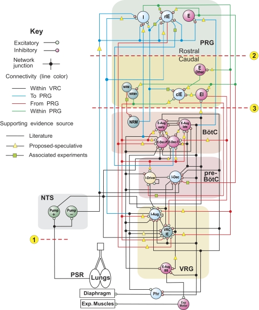
FIG. 1.
Schematic of the initial model of the brain stem respiratory network. To facilitate the tracing of pathways, regional connections are color-coded and dots are used to mark branch points of divergent projections. Both evidence-based (see text) and more speculative functional connections are represented in the model (see key). Model parameters for cell properties and connections are detailed in Tables A1–A4 of the appendix. Circled numbers and dashed lines in this and subsequent model diagrams label specific simulated perturbations applied. See text for details.

