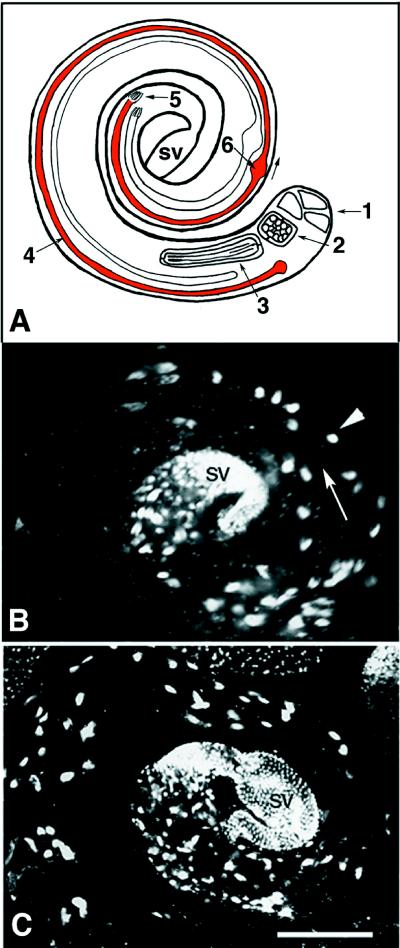
Figure 5.
Testis morphology in 95F myosin mutants. Schematic diagram of single wild-type testis indicates the key features in maturation of the male germ cells. Drosophila testes appear as spiral structures. The elongated spermatids are oriented with the caudal end of the spermatid tails toward the apical end and the spermatid heads toward the seminal vesicle (sv; terminal end). A single cyst of 64 spermatids undergoing individualization is shaded red. (1) Hub of stem cells at the apical end from which the germ cells and cyst cells are derived. (2) Cyst of 64 haploid germ cells before elongation. (3) Cyst of 64 haploid germ cells undergoing elongation, interconnected by cytoplasmic bridges. (4) Single cyst, which has undergoneelongation (red) and is in the process of individualizing. The IC moves in the direction of the arrow. (5) Terminal end of the cyst where the sperm and elongating spermatid nuclei reside. (6) Cystic bulge of the individualizing spermatids. Wild-type (B) and mutant (C) testes stained with DAPI to visualize DNA have similar overall morphology. Note that the region corresponding to the apical portion in which stem cell divisions are occurring is not shown in the micrographs. Large spots of DAPI staining material are bundles of nuclei of either individualized sperm or spermatids that have yet to complete individualization. A single bundle of 64 spermatids (arrowhead) is indicated. In wild type (B) the nuclear clusters are localized to the terminal third of the testes, whereas in the mutant (C) the nuclear clusters are present more toward the apical end of the testes. The arrow indicates the direction in which the ICs travel during individualization. Single bundles are shown in higher magnification in Figures 6 and 7. sv, seminal vesicle. Bar, 10 μm.

