Fig. 4.
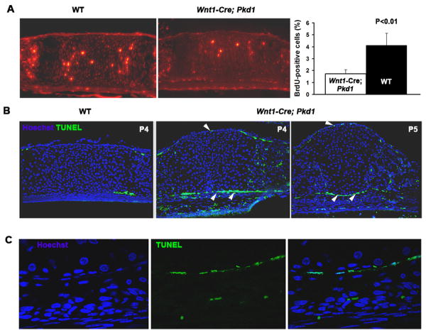
Pkd1-deficient presphenoid synchondrosis is characterized by a reduced proliferation of chondrocytes and increased apoptosis of the perichondrial cells. (A) Representative example of BrdU-specific staining of P3 presphenoid synchondrosis in control and Wnt1-Cre;Pkd1 cranial base (left and middle panels). Proliferative rate of control (black bar) and Pkd1-deficient (white bar) PSS chondrocytes is shown as average percent of BrdU-positive cells relative to total cell population (n=5). All error bars are s.d., P<0.01, student t test (right panel). (B) TUNEL staining of the control and Pkd1-deficient presphenoid synchondrosis prior to its obliteration reveals cell death within the perichondrium of the mutant (arrowheads). (C) High magnification of the single layer of apoptotic cells in the perichondrium of the mutant PSS as its closure proceeds.

