Fig. 6.
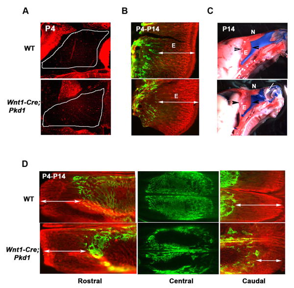
Wnt1-Cre;Pkd1 mice display normal proliferation and endochondral ossification of nasal septum, but reveal retarded growth of the caudal nasal bone. (A) Phosphorylated histone 3 labelling of the nasal cartilage (outlined) demonstrates a comparable level of interstitial chondrocytic proliferation in the mutant and control pups. (B) In vivo calcein and alizarin complexon double-labelling of bone tissue deposited on postnatal day 4 (green) and day 14 (red) demonstrates that the endochondral growth of the ethmoid bone is not affected by Pkd1 inactivation, as its mineralization rate (double-headed arrows) is similar in the Wnt1-Cre;Pkd1 and control littermates. (C) Impaired nasal bone growth in the Pkd1 knockout mice results in lateral dislocation of the nasal cartilage (arrow). Arrowheads mark the limits of the ethmoid bone. (D) In vivo calcein/alizarin double-labelling of bone deposited on postnatal day 4 (green) and day 14 (red) shows normal growth of the mutant and wild type rostral nasal bone (left panels, double-headed arrows). In contrast, mineralization of the caudal nasal bone is markedly reduced in the Pkd1-deficient mice relative to control (right panels, double-headed arrows). Note a delay in postnatal intramembranous ossification evident in the nasal mutant bone relative to control (central panels). E-ethmoid bone, N-nasal bone

