
Figure 2. CP increases the rate of Arp2/3 nucleation during steady-state motility.
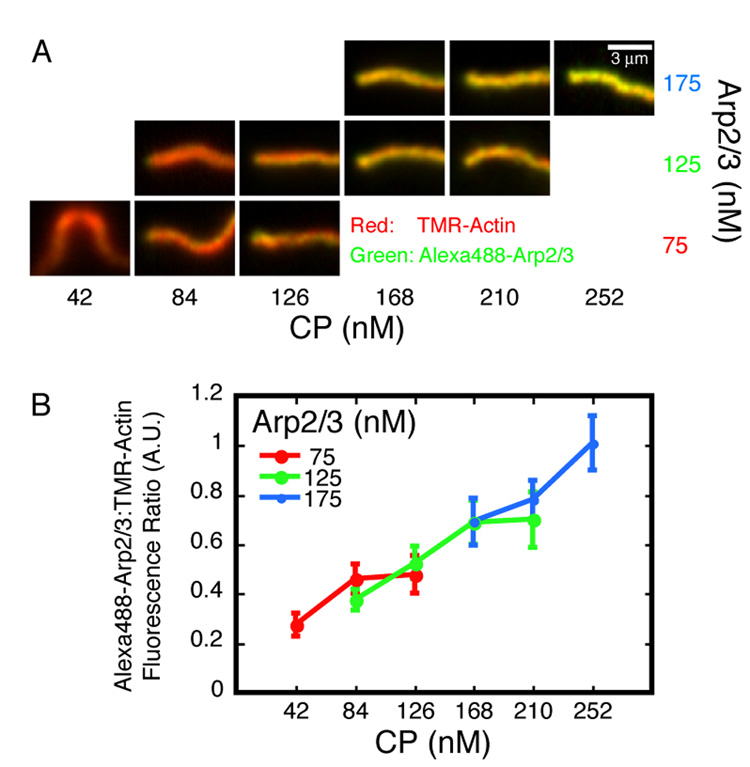
A. Representative actin comet tails assembled on 220 nm ActA-coated beads in the presence of various concentrations of Arp2/3 (10% Alexa488-labeled) and CP as indicated. Images were acquired approximately 30 minutes after mixing. Tails are framed with the beads (not shown) to the left of each panel, except the lower left panel (75 nM Arp2/3, 42 nM CP) where the bead would be top-center. This condition, as well as lower [CP] than that shown for the higher two Arp2/3 concentrations, produces twin tails on every bead. The red (i.e. actin) channel of each image was set to 90% saturation; this value was scaled by the same factor to determine the green (i.e. Arp2/3) channel display limits for all images. Conditions: 7.5 µM actin (5% TMR-labeled), 4 µM cofilin, and 3 µM profilin.
B. Fluorescence intensities along single-pixel spines of actin comet tails were measured. Each data point is the ratio of Alexa488-Arp2/3 to TMR-actin intensity averaged over the first micron length of the tails. Error bars are standard deviations (average n=27).