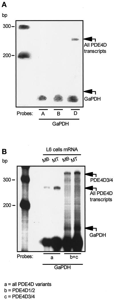
Figure 4.
RNase protection analysis of the PDE4D genes expressed in L6-C5 myoblasts and myotubes. L6-C5 cells were cultured for 24 h in complete medium and then shifted to serum-free medium in the absence (myoblasts, MB) or presence of 0.1 μM AVP (myotubes, MT). After 5 d, Poly(A)+ RNA was extracted, and RPA was performed as detailed in MATERIALS AND METHODS. Autoradiographies of the PAGE gels of protected fragments are shown. (A) The experiment was performed on myoblast mRNA with one of three probes recognizing all the products of PDE4A (expected size of protected fragment, 215 bp), PDE4B (expected size of protected fragment, 153 bp), and PDE4D genes (size of the protected fragment, 270 bp). mRNA integrity was assessed by glyceraldehyde-3-phosphate dehydrogenase–protected fragments (size, 130 bp). (B) The experiment was performed with either a probe recognizing a region common to all PDE4D transcripts (a) or a mixture of probes recognizing, respectively, the 5′ end of PDE4D1 and PDE4D2 transcripts (expected sizes of protected fragments, respectively, 293 and 170 bp) (b) and the 5′ end of PDE4D3 and PDE4D4 transcripts (size of protected fragment, 350 bp) (c). The size of the major band indicates that a fragment corresponding to PDE4D3/4 was protected, whereas no band corresponding to the size of the PDE4D1/2-specific probe could be detected.

