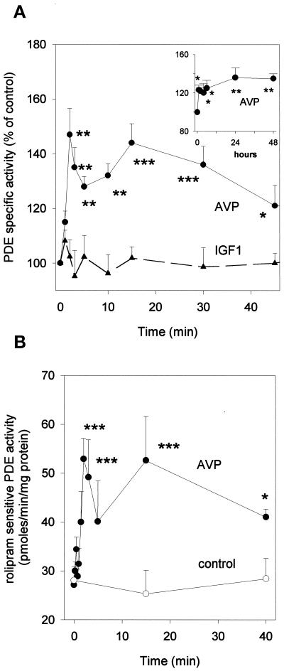
Figure 6.
PDE4 activity is stimulated by AVP-treatment of L6-C5 cells. (A) Effects of AVP and IGF1 on total cAMP-PDE activity of L6-C5 cells. AVP (1 μM), IGF1 (1 nM), or saline (control) was added at time 0 to L6-C5 myoblasts, which had been cultured for 2 d in serum-free medium. Cells were harvested at the indicated times (an extended time course up to 48 h is shown in the inset), and immediately homogenized and assayed for total cAMP-PDE activity. Data are means ± SEM for nine or more samples in at least five different experiments for AVP (●) and five or more samples in three different experiments for IGF1 treatment (▴). (B) Effect of AVP treatment on PDE4 activity of L6-C5 cells. Cells were treated with 1 μM AVP ([●) or left untreated (○) for the indicated times. They were then rapidly homogenized and assayed for cAMP-PDE activity, in the presence and absence of 10 μM rolipram. PDE4 activity was evaluated as the rolipram-inhibited fraction of PDE activity. Data are means ± SEM for n = 5–7 samples from four different experiments. *, Significance at p < 0.05; **, p < 0.02; ***, p < 0.01.

